COLOSTRUM
SUMMARY OF PRODUCT CHARACTERISTICS
The increasing demand for colostrum is driven not merely by the rise in research studies, but primarily by the enduring fascination with its live cell phenomenon. This is because the immune and growth factors crucial for newborns also offer significant benefits for adults. The immune protection passed on to infants, known as loan immunity, is a critical aspect that cannot be overlooked.
Unlike humans, where maternal antibodies are transferred to the fetus via the placenta and umbilical cord, thereby neutralizing microbes from birth, this mechanism is absent in mammals like cows and horses. Consequently, newborn calves and foals depend entirely on their mother’s initial milk, colostrum, which contains a full spectrum of immunoglobulins. Insufficient absorption of these immunoglobulins is linked to higher rates of illness and death among newborn animals within the first few weeks.
Bovine colostrum (BC) contains significantly higher concentrations of bioactive substances compared to human colostrum. For instance, BC has about 20 times more protein and at least 10 times higher concentrations of IgG (immunoglobulin G).
Given its rich antibody content, it is not surprising that naturopath Christoph Hufeland recognized colostrum as a universal remedy as early as the late 18th century. The influence of American Civil War reports on its use as a natural antibiotic for wound care may have further spurred its reputation. By the 1950s, it became evident that BC offered therapeutic benefits beyond immunoglobulins, showing efficacy in treating conditions such as chronic polyarthritis and even improving hay fever, highlighting its potent immunomodulatory properties.
BIOLOGICALLY
ACTIVE SUBSTANCES
Since the cow‘s originally viscous and yellow first milk (beestings) also serves as an energy and food source for normal metabolic processes and the growth of the calf, this means that it contains at least 90 biologically active substances (8). They comprise proteins, fat and lactose as well as antimicrobial factors, immunostimulant peptides and several important growth factors. We describe below only those active substances that are of importance when consciously selecting important of possible areas of application.
IMMUNE FACTORS
OF BOVINE COLOSTRUM
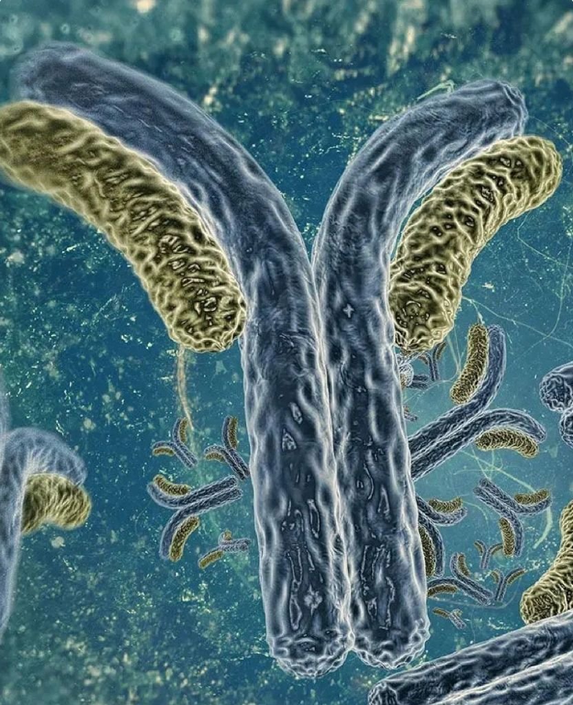
IMMUNOGLOBULINS
BBC contains the immunoglobulins IgG (IgG1/IgG2), IgM, IgA, IgE and IgD. Within 24 hours after calving, 90% of BC consist of IgG9, three-fourths of which, in turn, are made up exclusively of IgG1 (46.4 mg/mL) (10). They are then followed by IgM (6.8 mg/mL) and IgA (5.4 mg/mL). Immunoglobulins thereby make up 70%-80% of the total protein content of colostrum (11).
Externally sourced immunoglobulins work exactly like antibodies formed by the body itself, reacting to antigens, namely the surface molecules of bacteria or glycoproteins of viruses, for example. This occurs based on a kind of lock-and-key principle where the antibody fits precisely into the antigen matching it. This usually leads to the clumping of pathogens, preventing them from penetrating into a cell, for example. At the same time, they become marked so that Tcells of the immune system as well as phagocytes can start working on their actual elimination. The so-called complement-mediated bacteriolytic reaction is one of the most important functions of Ig antibodies. Contributing to this is the fact that the complete complement system is found in BC.
Immunoglobulins thus provide protection from bacteria, viruses, fungi and parasites. In an investigation of antibody-mediated antimicrobial activity, all BC samples from normal healthy cattle had a natural bactericidal effect on Helicobacter pylori (12). Raw, that is, unpasteurized milk or BC from non-immunized cows contains antibodies against the human rotavirus, for example (13). BC also contains specific Ig against lipopolysaccharides of the dominant organisms Salmonella enteritidis, S. typhimurium and Shigella flexneri (14). Natural antibodies against the colonization antigen (CFA1) of human enterotoxigenic E. coli were also found in normal BC (15).
GROWTH FACTORS
BC contains a series of important growth factors that can promote the growth, maturation and repair of bones, muscles, nerves, connective tissue, cartilage, skin tissue and modulate the gastrointestinal mucous membrane (61). Growth factors are relatively stable against heat and acidic conditions. They therefore survive harsh digestive processes as well as gastric acid (62).
INSULIN-LIKE GROWTH FACTOR 1 AND 2 (IGF-1/-2)
The IGF-1 concentration in BC is 200-2,000 μg/L and hence about 10 times greater than in human blood serum (63). The amount of IGF- 2 is 1,800 μg/L64. Bovine IGF, in terms of the first 30 amino acids, is completely homologous to human IGF and hence has an identical effect (65). With regard to its intestinal impact potential, IGF-1 primarily stimulates the cell proliferation of intestinal crypts, while IGF-2 controls the mechanism of intestinal epithelial cells (66).
TRANSFORMING GROWTH FACTOR (TGF-ALPHA)
This growth factor in BC is already naturally synthesized in the mucous membrane of the gastrointestinal tract as well (67). Systemic administration of TGF-alpha stimulates the secretion of mucus, cell growth and wound healing, and reduces acid formation. Its physiological role is cellular differentiation and migration, and it is used for the repair and maintenance of intestinal epithelial integrity (68).
TRANSFORMING GROWTH FACTOR (TGF-ß)
The TGF-ß2 concentration in BC is 150-1,150 ng/mL69. This concentration is enough to prevent indomethacin-caused gastric lesions in rats, for example (70). It resembles an “attractant” that has a powerful chemical attraction to neutrophils. It plays an important role in repair processes by promoting the migration of epithelial cells to injured, empty areas in order to restore epithelial continuity (71).
PLATELET-DERIVED GROWTH-FACTOR (PDGF)
This growth factor, also contained in BC, is normally produced by platelets and macrophages. PDGF is a potent mitogen for fibroblasts. In particular, it facilitates wound healing in ulcers, for example after prolonged NSAID use (72).
BIOAVAILABILITY OF
IMMUNOGLOBULINS
In-vitro studies have shown that BC is relatively resistant to proteolysis by digestive enzymes and also survives gastric acid unscathed (16,17,18). In the human digestive tract, bovine IgG is broken down by the proteases pepsin, trypsin, chymotrypsin, carboxypeptidase and elastase into the antibody fragments F(ab)2, Fab and Fc, for example. This, however, has no harmful effect because the fragments F(ab)2 and Fc contain the neutralizing effect of an intact IgG molecule.
CYTOKINES
Cytokines are regulatory proteins (peptides) that serve to control immune response. They are normally produced in the human body by macrophages, B lymphocytes, natural killer cells (NK) and fibroblasts. Above all, they are also essential messengers for cellular communication (19). Cytokines found in BC are IL-1ß, IL-6, TNF alpha, interferon gamma and IL-1 receptor antagonists. They stimulate the immune system and are involved in inflammatory processes. Cytokines also act in conjunction with other defense components present in BC such as immunoglobulins, lactoferrin and lactoperoxidase
Little is known about the cytokine content of commercially processed BC. However, there are studies on the extent to which BC supplementation stimulates the body’s own cytokine production. IFN-gamma, IL-10 and IL-2 secretion are shown in vitro to increase linearly with BC concentration. In cells co-cultured with lipopolysaccharides, the secretion of TNF alpha, IL-6 and IL-4 decreased. This suggests that BC supplementation reduces pro- inflammatory cytokine production after strenuous exercise (21) as this is typically associated with a high lipopolysaccharide level.
LACTOFERRIN
In BC, lactoferrin and lactoperoxidase are among the dominant and best studied antimicrobial components that are effective against all types of microorganisms, at least in vitro. Even small quantities can have an enormous biological effect. Significantly, lactoferrin is found in almost all body fluids such as tears22, saliva, sweat, vaginal secretion23 as well as nose and bronchial secretion24. Lactoferrin (racedependent/24 hours after calving) is present in BC in a concentration between 2 mg/mL and 0.5 mg/mL25.
Lactoferrin owes its bacteriostatic effect primarily from its extremely strong iron-binding capacity (26). It acts by removing iron, which bacteria need to live. Notwithstanding the above, lactoferrin also has an ironindependent bactericidal property since it increases the membrane permeability of microbes, leading to their death. Lactoferrin is also effective against viruses27, fungi and protozoa. It has a very strong effect on HIV28, polio (29) and herpes (HSV)30, for example. However, this is not the same effect as that of PRPs, which inhibit viral replication, but it acts rather by binding directly to virus particles31. It also appears to have a modulating effect on intestinal microbes (32), and even amplifies the proliferation and differentiation of intestinal epithelial cells (33). This still needs to be studied more closely with respect to the microbiome area of application as well as NSAID and antibiotic injuries.
Lactoferrin also influences immune functions like cytokine synthesis and the maturation, migration and activation of macrophages, granulocytes, natural killer cells (NK) as well as T and B cells by occupying their specific receptors (34).
GANGLIOSIDES
The gangliosides present in BC promote the proliferation, maturation and activation of lymphocytes, dendritic cells, cytokine production and intestinal IgA secretion (43). In particular, ganglioside 3 (GD3) functions as a false receptor for toxins and adhesins of pathogenic bacteria (44). Gangliosides also promote the proliferation of useful microbiota like bifidobacteria and thus participate indirectly in the body’s own immune defense45.
PROLINE- RICH POLYPEPTIDES (PRPs)/COLOSTRININ
The proline-rich polypeptides (PRPs) present in microorganisms, plants and animals (a conglomerate of at least 32 different peptides) play an important role in biological processes. In this way peptides Herpes viruses (47,48,49), Epstein-Barr viruses (50), HIV51 and the measles virus (52) prevent their replication.
In the 1970s PRPs were isolated for the first time from the colostrum of sheep and were then called colostrinin (CLN). In the meantime, it turned out that bovine colostrum (BC) contains double the amount of PRPs compared to the original substances derived from the first sheep’s milk (53).
LACTOPEROXIDASE (LPO)
The biological function of LPO mainly involves defense against bacterial infections. The bacteriostatic and bactericidal potential needed here applies to a wide spectrum of microorganisms35, especially to gram-negative organisms such as pseudomonas, coliforms, salmonella and shigella. LPO also has an antiviral (e.g. HIV) 36 and tumoricidal (37) effect, although the latter has only been confirmed in vitro to date.
OLIGOSACCHARIDES
In addition to lactose, which is the main type of sugar, BC contains a series of sugar types such as glycolipids, glycoproteins, cellulose, glycosaminoglycan and mucin (38,39). They function as “false” receptors and thus prevent the binding of bacteria and viruses to intestinal cells (40). BC oligosaccharides also activate typical toll-like receptors (TLR-2 and TLR-4) of the intestinal epithelium, leading to an increase in the cytotoxicity of natural killer cells (NK) (41).
In anticipation of the microbiome area of application, discussed later, it is worth emphasizing that some sugars like fructo- oligosaccharides and galacto-oligosaccharides act as prebiotics since they promote the growth of a healthy intestinal microbiome such as bifidobacteria and lactobacilli (42).
CLN binds to receptors of immune cells such as B cells from bone marrow and thus causes quicker maturation. Overall, there are indications for counting PRPs/CLN simply among the class of cytokines known as “super cytokines” (54) which means they are produced not just by the immune system but by the mammary glands. In any event, they can stimulate both the production of leukocytes and NKs55 as well as the formation of other cytokines such as those of interferon, TNF in human leukocytes or a blood culture. In an in vitro study, PRPs stimulated IFN and TNF production in peritoneal cells of mice 30-fold (56).
It is also surprising that PRPs significantly reduce the production of IgE/IgG1, the eosinophilia of the respiratory tract, mucus production and even hypersensitivity; in other words, the typical symptoms caused by allergens like ragweed pollen or household dust mites. From this one can draw the conclusion that PRPs can prevent all types of allergic reactions (57).
And then there is evidence from studies that PRPs can stimulate neuronal growth, suppress chronic microglial production and even reduce beta-amyloids (58,59,60). This will be discussed in the Alzheimer’s disease area of application.
OTHER INGREDIENTS
NUKLEOTIDES
Nucleotides present in BC act on the integrity of the intestinal mucosa and the commensal flora (microbiome). Apart from this, they affect the proliferation potential of growth factors (73).
LYSOZYME
This BC enzyme has antimicrobial properties against gram-negative and gram-positive bacteria since it can break up the peptidoglycan layer of bacterial cells (74).
MICRO RNA AND MESSENGER RNA
BC also contains microRNA and mRNA. Due to their transport in microvesicles they appear to be resistant to gastric acid and RNase development of the gastrointestinal tract and the immune system its exact function is not quite clear yet.
ANTIOXIDANTS
BC contains a wide range of enzymatic and non-enzymatic components that can have an antioxidant and, in rare cases, a prooxidant effect depending on physiological state (77). The enzymatic antioxidants of BC include lactoperoxidase, catalase, superoxide dismutase and glutathione peroxidase. The non-enzymatics include vitamins E, A and C, lactoferrin, selenium, copper, zinc and cysteine78. They easily stand up to any comparison with proven antioxidant compounds.
REFERENCES
1. Goldman AS. Pediatr Infect Dis J 1993; 12: 664-674
2. McGuire TC et al. J Am Vet Med Assoc 1976; 169: 713-8
3. Rea DE et al. J Am Vet Assoc 1996; 208: 2047-9
4. Carillo AE, Koutedakis Y, Flouris AD. Cell Mol Biol (Noisy-le-grand). 2013; 59(1): 84-88
5. Christoph Wilhelm Hufeland, Hrsg: Bibliothek der practischen Heilkunde (Journal der praktischen Arzneykunde und Wundarzneykunst. 1795-1836 (83 Bände)
6. Struss H. A review. J Immun Milk 1964; 1: 23-42 ^4e^^23
7. Campbell B, Petersen WE. Diary Sci 1963; 25(9): 345-58
8. Kehoe SI, Jyarao BM, Heinrichs AJ. J Diary Sci 2007; 90(9): 4108-16
9. Alexieva B, Markova T, Nikolova E. Czech J Food Sci 2004; 22(1): 73-79
10. Gapper LW et al. Review. Anal Bioanal Chem 2007; 839(1): 93-109
11. Larson BL. In: Fox P.F. editor. Advanced Daily Chemistry Proteins. 2nd. Elsevier Applied Science. London, UK. 1992
12. Korhonen H et al. J Appl Bacteriol 1995; 78(6): 655-62
13. Yolken RH et al. N Engl J Med 1985; 312: 605-610
14. Losso JN et al. Comp Biochem Physiol B 1993; 106(4): 919-23
15. Facon M, Skura BJ, Nakai S. Food Res Inter 1995; 28: 387-396
16. DeRham O, Isliker H. Int Arch Allergy Appl 1977; Immunol. 55: 61-69
17. Brock JH, Pineiro A, Lampreave F. Ann Rech Vet 1978; 9(2): 287-94
18. McLead RE, Gregory SA, Infect Immun 1984; 44(2): 474-8
19. Garofallo R. J Pediatr 2010; 156 (Suppl 2): 36-40
20. Shing CM et al. J Interferon Cytokine Res 2008; 29(1): 37-44
21. Jeukendrup AE et al. Clin Sci (London) 2000; 98: 47-55
22. Park JH et al. Exper Dermatol 2011; 30(4): 369-371
23. Cohen MS et al. Am J Obstetr Gynecol 1987; 157(5): 1122-25
24. Elrod KC et al. Am J Respir Critic Care Med 1997; 156: 375-81
25. Tsuji S et al. J Dairy Sci 1990; 73(1): 125-8
26. Sanchez L, Calvo M, Brock JH. Arch Dis Child 1992;67(5): 657-61
27. Harmsen MC et al. J Infectious Diseases 1995; 172: 380-88
28. Harmsen MC et al. J Infect Dis 1995; 172(2): 380-388
29. Superti F et al. Med Microbiol Immunol 1997; 186(2-3): 83-91
30. Hasegawa K et al. Jpn J Med Sci Biol 1994; 47(2): 73-85
31. Van der Strate BW et al. Antiviral Res 2001; 52(3): 225-39.
32. Anand N et al. Drug Des Devel Ther 2015; 9: 821-35
33. Buccigrossi V et al. Pediatr Res 2007; 61(4): 410-4
34. Legrand D et al. Cell Nol Life Sci 2005; 62(22): 2549-59
35. Wolfson LM, Sumner SS. J Food Protect 1993; 56: 887-92
36. Courtois B et al. J Biologie Buccale. 1990; 18: 71-74
37. Stanislawski M et al. Cancer Res 1989; 49: 5497-5504
38. Urashima T et al. Biosci Biotechnol Biochem 2013; 77(3): 455-66
39. Úrashima T, Fukuda K, Messer M. Animal 2012; 6(3): 369-74
40. Newburg DS. J Anim Sci 2009; 87(Suppl 13): 26-34
41. Wong EB et al. Nutr Res 2014; 34(4): 318-25
42. Tlaskalova-Hogenova H et al. Immunol Lett. 2004; 93(2-3): 97-108
43. Rueda R. Br J Nutr 2007; 98 (Suppl 1): 68-73
44. Idot T, Tawakami H. Biosci Biotech Biochem 1995; 59(1): 69-72
45. Nakano T, Sugawara M, Kawakami H. Acta Paediatr Taiwan 2001; 42(1): 11-17
46. Otvos L. J Pept Sci 2000; 6: 497-511
47. Pizza G et al. Biother 1994; 8(1): 63-68
48. Pizza G et al. Biother 1996; 9(1-3): 67-72
49. Meduri R et al. Biother 1996; 9(1-3): 61-66
50. Wieczorek Z et al. Arch Immunol Ther Exper (Warszava): 1989; 37(3-4): 313-322
51. Raise E et al. Biother 1996; 9(1-3): 49-54
52. Ferrer-Argote VE et al. In Vivo 1994; 8(4): 555-7
53. Sokolowska A et al. Internat Diary J 2008; 18: 204-209
54. Zablocka A et al. Eur Cytokine Netw 2001; 12(3): 462-7
55. Kruzel ML et al. J Mol Neurosci 2001; 17(3): 379-89
56. Blach-Olszewska Z, Janusz M. Arch Immunol Ther Exp /Warsz) 1997; 45(1): 43-7
57. Boldogh I et al. Int Arch Allergy Immunol 2008; 146(4): 298-306
58. Inglot AD et al. Arch Immunol Ther Exp (Warsz) 1996; 44(4): 215-24
59. Schuster D et al. Neuropeptides. 2005; 39(4): 419-26
60. Janusz M, Zablocka A. Curr Alzheimer Res 2010; 7(4): 323-33
61. Playford RJ et al. Clin Sci (Lond) 2001; 100(6): 627-33
62. Lowe WL. In: LeRoith D. ed. Insulin-like growth factors: molecular and
cellular aspects. Boca Raton, Fl. CRS Press 1991: 49-85
63. Mero A et al. J Appl Physiol (1985); 1997; 83(4): 1144-51
64. Vega JR et al. J Anim Sci 1991; 69(6): 2538-47
65. Red LC et al.Biochem J 1986; 233: 215-221
66. Jehle PM et al. Horm Metab Res 1999; 32(2-39): 97-102
67. Cartlidge SA, Elder JB. Cancer 1989; 60(5): 657-60
68. Koyama S, Podolsky DK. J Clin Invest 1989; 83(5): 1768-73
69. Pakkanen R. J Innunoassay 1998 Feb; 19(1): 23-37
70. Marchbank T, Playford RJ. Gut 1998; 42(Suppl): A68 (abstr.)
71. Nguyen DN et al. Am J Physiol Gastrointest Liver Physiol 2014; 307(7): 689-99
72. Shing W, Klagsbrunn M. Endocrinol 1984; 115(1): 273-82
73. Belo E et al. Gut 2006; 55(2): 165-71
74. Artym J, Zimecki M. Postpepy Med Dosw (Online) 2013; 67: 800-16
75. Izumi H et al. J Diary Sci 2012; 95: 4831-4841
76. Sun Q et al. Protein Cell 2013; 4(3): 197-210
77. Przylbylska J, Albera E, Kankofer M. Reprod Domest Anim 2007; 42(4): 402-9
78. Pandey NN et al. J Vet Med Animal Health 2011; 3(3): 31-35